One of the classic dilemmas dentists face is when they encounter an endo-perio lesion. While there is a lot that one can do to salvage the tooth and restore its form and function, following the right clinical signs and symptoms to accurately diagnose the lesion and take a step-wise approach to managing it is crucial for treatment success.
This month, we’ve got you a brief guide for identifying and managing endo-perio lesions in your practice!
| Type of Lesion | Clinical Characteristics |
|---|---|
| Primary Endodontic Lesion |
|
| Primary Periodontal Lesion |
|
| Primary Endodontic Lesion with Secondary Periodontal Involvement |
|
| Primary Periodontal Lesion with Secondary Endodontic Involvement |
|
| True Combined Lesion |
|
Clinical signs that will help you diagnose if the lesion is endodontal or periodontal in origin:
| Clinical Sign | Endodontal Lesion | Periodontal Lesion |
|---|---|---|
| Pulpal vitality | Non-vital | Vital |
| Plaque / Calculus | Unrelated | Primary cause |
| Inflammation | Acute | Chronic |
| Periapical lesion | Radiolucent | Usually not related |
| Vertical bone loss | Absent | Present |
| Junctional epithelium | No apical migration | Apical migration present |
| Gingiva | Normal | Recession |
| Bone loss | Wider apically | Wider coronally |
Once you have identified the type of lesion, here’s a simple flow that will help you decide on a suitable treatment plan:
Step 1: Perform a pulp vitality test
| Pulp Status | Likely Diagnosis |
|---|---|
| If the pulp is vital | You may have a periodontal lesion in hand |
| If the pulp is non-vital | You may be dealing with an endodontal lesion |
Step 2: Check for pockets
| Probing Depth | Likely Origin of Lesion |
|---|---|
| If the probing depth is < 3 mm | The lesion may be endodontic in origin |
| If the probing depth is > 3 mm | The lesion may be periodontal in origin |
| Prognosis | Recommended Management |
|---|---|
| If the prognosis is good | Perform endodontic therapy, and restore and review the tooth over the next few weeks and months. |
| If the prognosis is poor | Extract the tooth. |
| Prognosis | Recommended Management |
|---|---|
| If the prognosis is good | Perform non-surgical periodontal therapy and review. Some patients may require supportive periodontal therapy. |
| If the prognosis is poor | Extract the tooth. |
Perform endodontic therapy, followed by simple hygiene phase → Monitor the tooth and evaluate after 2-3 months
If needed, periodontal therapy can be performed
If the lesion is due to perforation→ Seal the perforation and keep under observation.
If there is a fracture → Manage it appropriately.
Endodontal therapy followed by palliative periodontal therapy. Evaluate after 2 to 3 months.
Check for periapical resolution.
If periapical lesion is resolved→ The tooth may be grade 1 mobile and may require splinting.
If the periapical lesion is not resolved → Treat for root damage, osseous defects and aberrant local morphology.
In case of poor prognosis at any step, extraction is the best solution.
In the case of pockets, treat them appropriately depending on pocket depth.
Colgate’s PerioGard range has been specially formulated to help manage gingival and periodontal concerns. It offers effective protection, promotes healing, and ensures optimal oral health for your patients.
Key Benefits:
PerioGard Mouthwash: This broad-spectrum antimicrobial rinse, with a low-staining chlorhexidine formula, reduces plaque and gingivitis and gives you long-lasting freshness.
PerioGard Toothpaste: Formulated with a unique combination of 2% Zinc citrate and Fluoride, it has high antibacterial efficacy to reduce plaque buildup and protect against gum problems.
PerioGard Gum ExpertToothbrush: It features ultra-soft, tapered bristles designed for gentle yet effective cleaning along the gum line and hard-to-reach areas.
References:
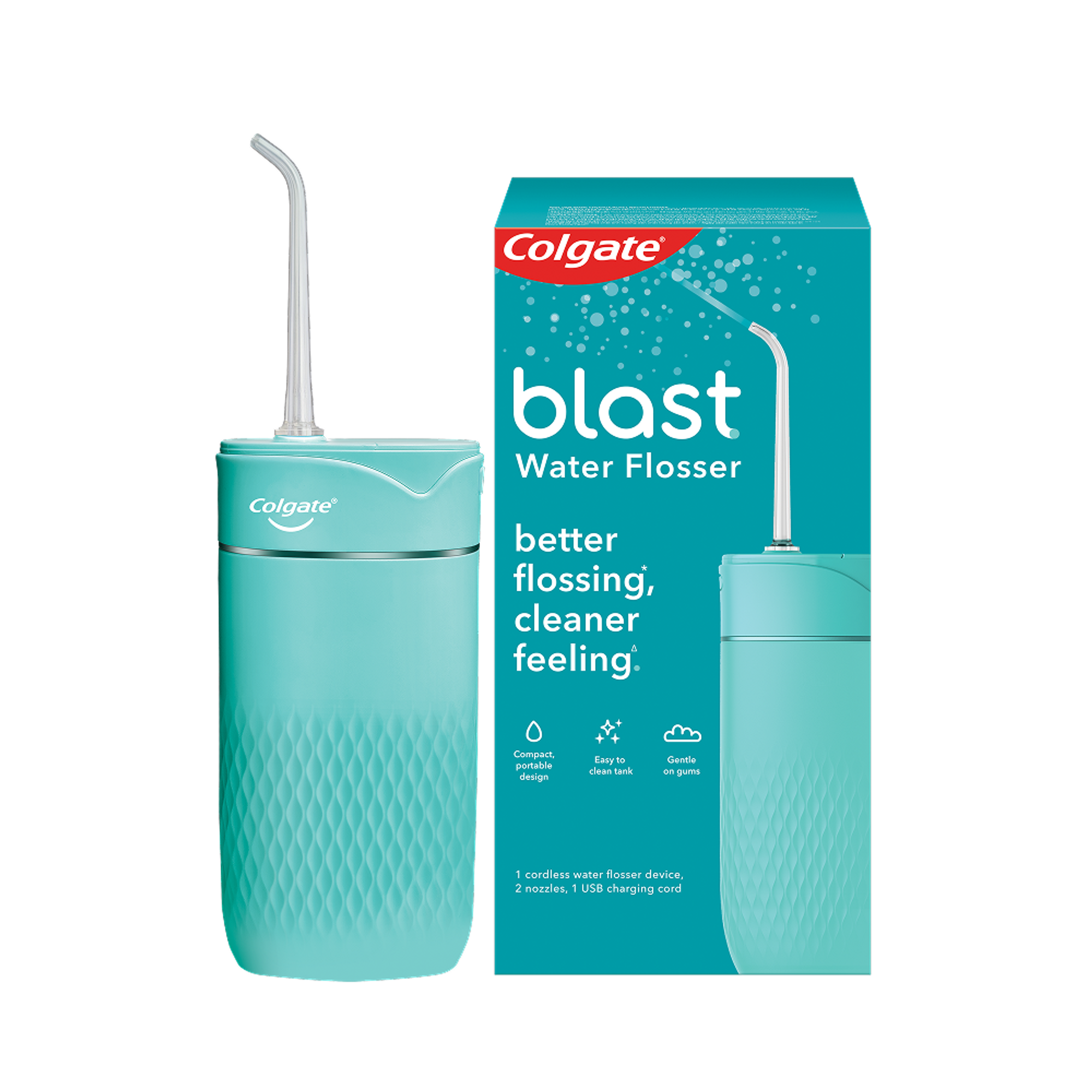
Colgate Water Flosser the fun and easy way for your patients to floss. Blasts away plaque between teeth and along the gums
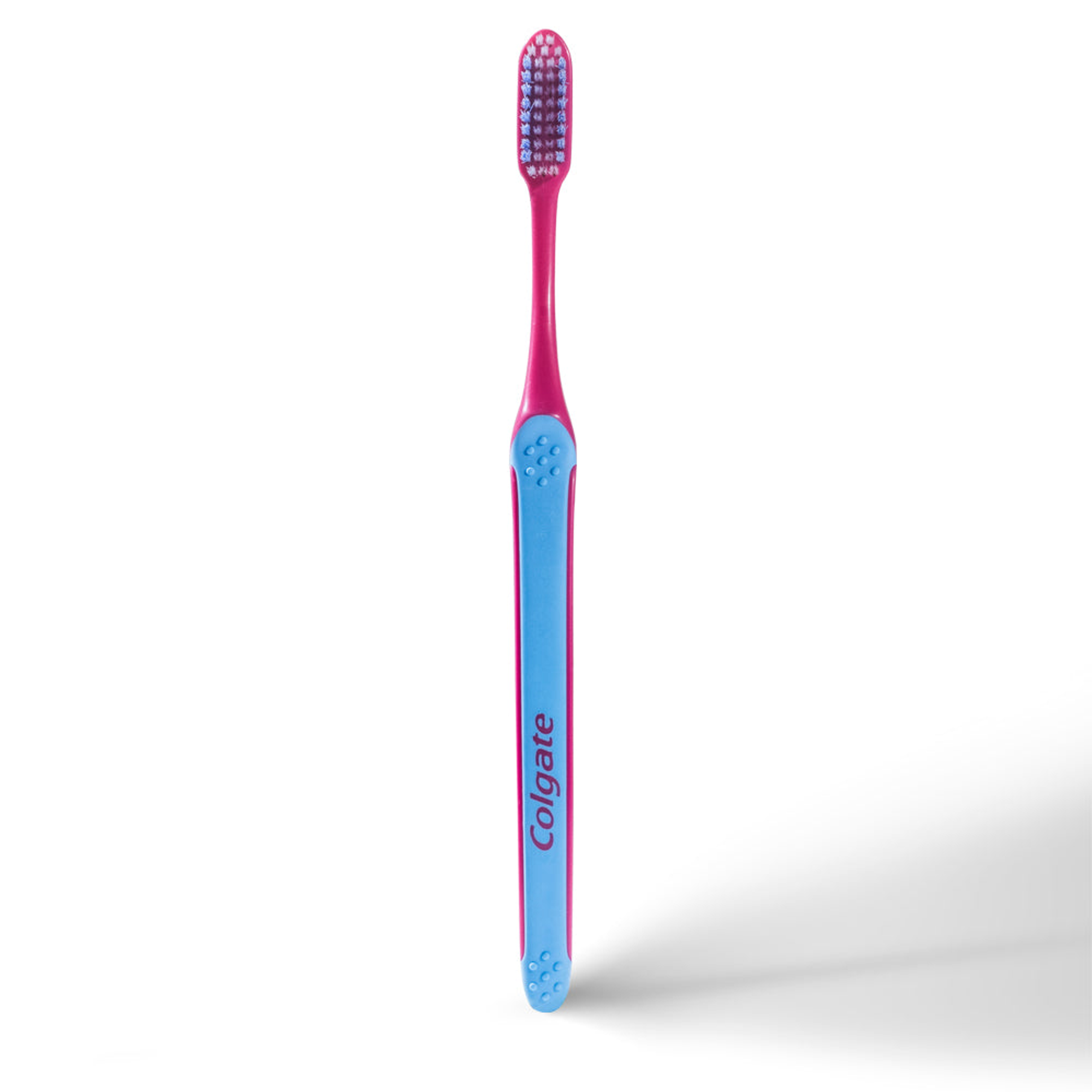
Slim Soft Orthodontic Toothbrush for patient who wear braces and need better plaque control